Doctoral Research at USC
Molecular imaging is a promising field that allows noninvasive in vivo visualization of specific molecular targets and pathways in living organisms. In recent years its sphere of influence has extended beyond early diagnosis of diseases as it has emerged as a valuable tool for drug discovery and development. Fluorescence molecular tomography (FMT) is a molecular imaging modality that exploits the specificity of fluorescent biomarkers, which absorb and emit light with a downward shift in the frequency. It generates volumetric functional images using near‐infrared light, which is safe and non‐ionizing, at a substantially lower cost than competing modalities like positron emission tomography (PET) and Single photon emission computed tomography (SPECT). While these very attractive features make this functional imaging modality well suited for preclinical research, the FMT problem is confounded by the high degrees of absorption and scattering of photons propagating through tissue, which make the inverse problem ill‐posed. The focus of my PhD research under the mentorship of Dr. Richard Leahy at the University of Southern California was to develop computational methods that enable us to reconstruct high resolution images of molecular targets in vivo in small animals and to develop a novel FMT system for small animal imaging.
Fast Forward Models
To accurately and efficiently predict the photon fluence on the animal surface, we have developed a fast perturbative approach based on the Born approximation to compute the system forward model with reasonable accuracy. Our method works for complex animal geometries and takes into consideration the heterogeneity of tissue optical properties. This work was accepted for oral presentation at the SPIE Medical Imaging Conference at San Diego in February, 2008.
Links: SPIE 2008 Paper
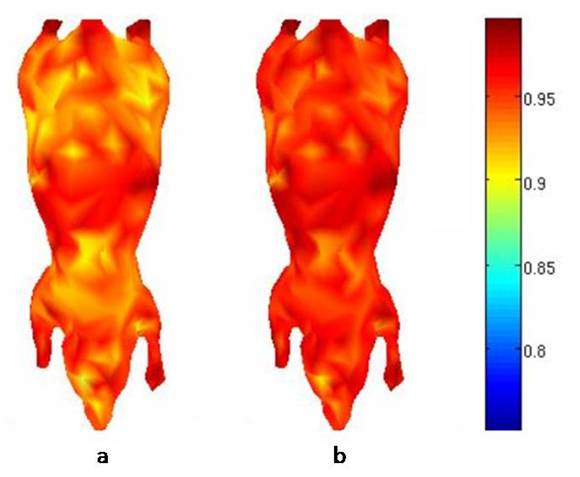
Correlation coefficients at surface surface detector nodes on the Digimouse atlas due to all the internal source points computed (a) for a homogeneous tissue model and (b) for our perturbative model using FEM as the reference. We observe that our approach gives better average correlation with FEM.
Optimal Illumination Patterns
We have also developed a systematic approach for designing efficient illumination patterns for FMT. Typically, multiple sets of data are acquired by illuminating the animal with different spatial patterns of near‐infrared light. However the choice of these patterns in most experimental setups is ad hoc and suboptimal. The key idea of our approach is to parameterize the illumination patterns and to formulate a constrained optimization problem that seeks to improve the conditioning of the Fisher information matrix for the FMT forward model. This work was selected for oral presentation at the IEEE Symposium on Biomedical Imaging at Boston in July, 2009, and was published in the journal Physics in Medicine and Biology.
Links: ISBI 2009 Paper, PMB 2010 Paper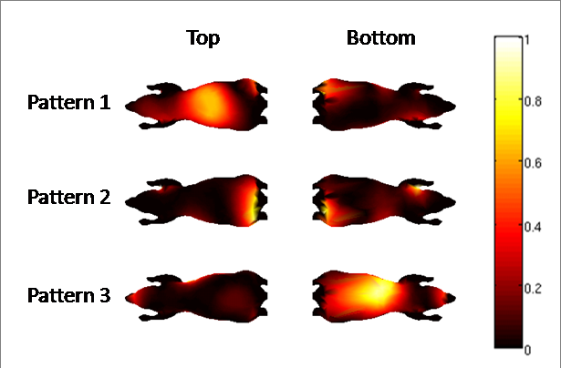
Projections of the top and bottom surfaces of the Digimouse atlas showing a set of 3 optimal patterns for fluorescence tomography.
Imaging System Development
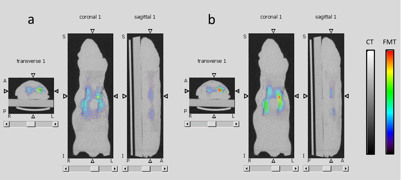
In collaboration with Prof. Simon Cherry's lab at UC Davis, we have built a prototype imaging system that uses a CCD camera for collecting multispectral fluorescence data and a compact conical mirror to provide a view of the entire surface of a mouse sitting on a transparent stage. We have demonstrated that the conical mirror geometry offers higher light collection efficiency and have developed a calibrated mapping model to extract surface fluorescence data from the mirror image. We have successfully used our system to conduct in vivo experiments. We published these results in the journal Optics Express in April, 2009. Our paper was selected to appear in the May issue of the Virtual Journal of Biomedical Optics.
Link: Optics Express Paper
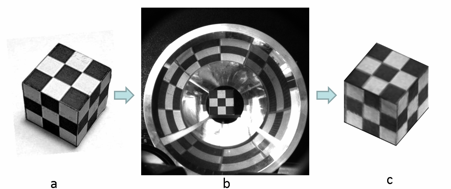
Conical mirror system calibration using a checkered cube: (a) Photograph of the original checkered cube, (b) CCD camera image, and (c) digital rendering of the cube with the reconstructed checkerboard pattern.
We have developed a second FMT imaging system in a three-way collaboration with Prof. Cherry's Lab and Cambridge Research and Instrumentation (CRi) (now a subsidiary of PerkinElmer). This system uses a pyramidal mirror setup for multi-view data acquisition and generates computer-controlled spatial patterns of illumination using a Texas Instruments DLP® projector. A set of preliminary phantom studies performed using this was presented at the OSA BIOMED Meeting at Miami in April, 2010.
Link: BIOMED 2010 Paper
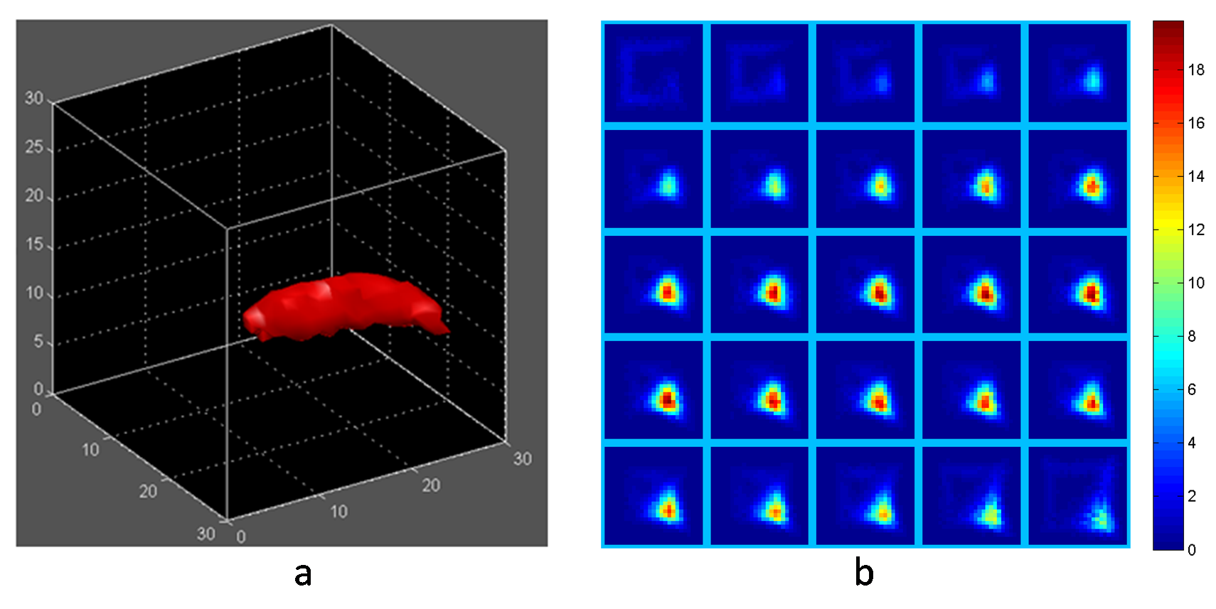
Phantom experiment using the pyramidal mirror setup: Reconstructed image of a fluorescent line source embedded inside a cubic tissue phantom displayed as (a) an isosurface using a 99th percentile intensity threshold for the image volume and (b) transverse slices through the axis of the fluorescent tube.
DigiWarp
We developed a three-step procedure named DigiWarp to warp the Digimouse atlas (a realistic mouse atlas based on CT and cryosection data of a male nude mouse) to fit the surface profile data. This method preserves organ information in the mouse tessellation thus allowing us to account for the optical inhomogeneity of animal tissue in our forward model. This work was also selected for oral presentation at the IEEE Symposium on Biomedical Imaging at Boston in July, 2009, and subsequently published in Physics in Medicine and Biology.
Links: ISBI 2009 Paper, PMB 2010 Paper
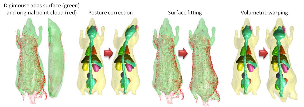
The DigiWarp procedure involving posture correction, surface fitting by minimizing asymmetric L2 distances, and finally volumetric warping by minimizing elastic energy.
Sparsifying and Smoothing Priors
Owing to excessive absorption and scattering of light through tissue, fluorescence tomographic image reconstruction is inherently ill-conditioned. A suitable regularizing penalty function that restricts the search space to a specific solution set is essential to facilitate reconstruction. Typically, fluorescent probes are locally concentrated within specific areas of interest (e.g., inside tumors). The commonly used L2 norm penalty generates the minimum energy solution, which tends to be spread out in space. Instead, we have developed an approach involving a combination of the L1 and total variation (TV) norm penalties, the former to suppress spurious background signals and enforce sparsity and the latter to preserve local smoothness and piecewise constancy in the reconstructed images. Our article describing this work was published as a Featured Article in Physics in Medicine and Biology.
Link: PMB 2012 Paper
Transverse, coronal, and sagital views from fused CT and FMT reconstructed images of a mouse-shaped phantom with two embedded fluorescent tubes. The FMT images were generated using (a) the traditional L2 penalty and (b) joint L1 and TV penalties which simultaneously enforce sparsity and smoothness.
In Vivo Experiments
We performed several preliminary in vivo experiments using the conical mirror setup and several of the computational tools described above. Shown below is the reconstruction result from an FMT study performed on a nude mouse with a xenograft melanoma (DX-6) dorsally located on the right flank. The reconstructed image shows fluorescence from the tumor bulge, some fluorescence from dye accumulating in the bladder, and some skin autofluorescence.
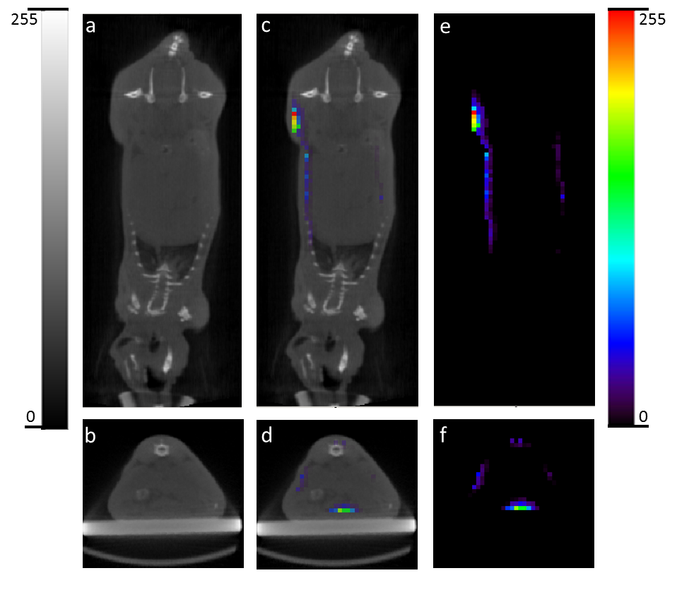
(a) Coronal and (b) transverse sections of a CT image of the mouse showing the tumor bulging on the right flank. (c) Coronal and (d) transverse overlay of CT and FMT images. (e) Coronal and (f) transverse sections of the FMT image showing the tumor on the right flank and fluorescence from the bladder.